Microscoop Mint is a novel cutting edge technology which allows for unprecedented precision in detecting proteomes at the subcellular level using microscopy-guided two-photon laser biotinylation. This platform was developed by Syncell and installed at the Radboudumc in the summer of 2025, making us one of the first labs in Europe to make use of this tool.
Why is this particularly valuable for cilia reseach?
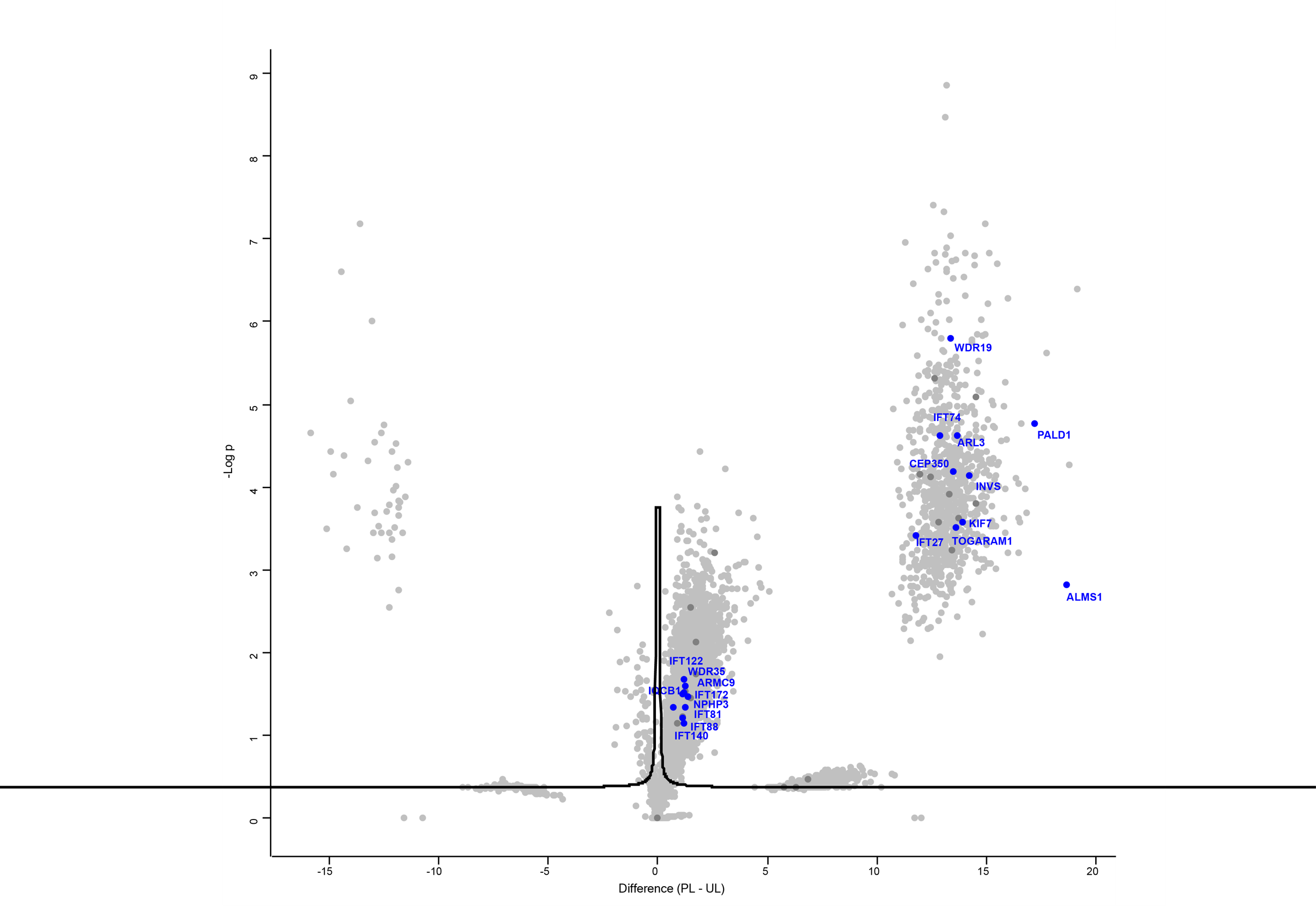
Defining the ciliary proteome in health and disease has remained a major challenge limited by the small size of the cilium and the lack of methods to isolate it from the rest of the cell. The ciliary membrane is continuous with the plasma membrane of the cell body and attempts to mechanically or chemically sever the two have remained inconsistent. Recombinant proteins, relying on the overexpression of cilia-targeted proximity labeling enzymes, while extremely valuable, are still time-consuming and limited to cell types which are highly transfectable. The Microscoop system resolves both issues, while further allowing us to also explore the ciliary proteome in-situ in tissues of interest, such as the retina, the kidney and the brain.
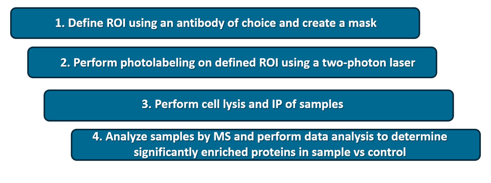
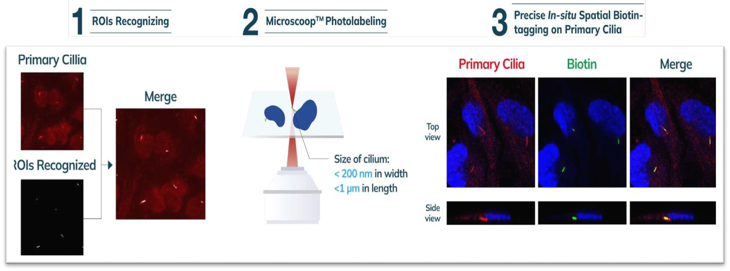
How does Microscoop work in a nutshell?
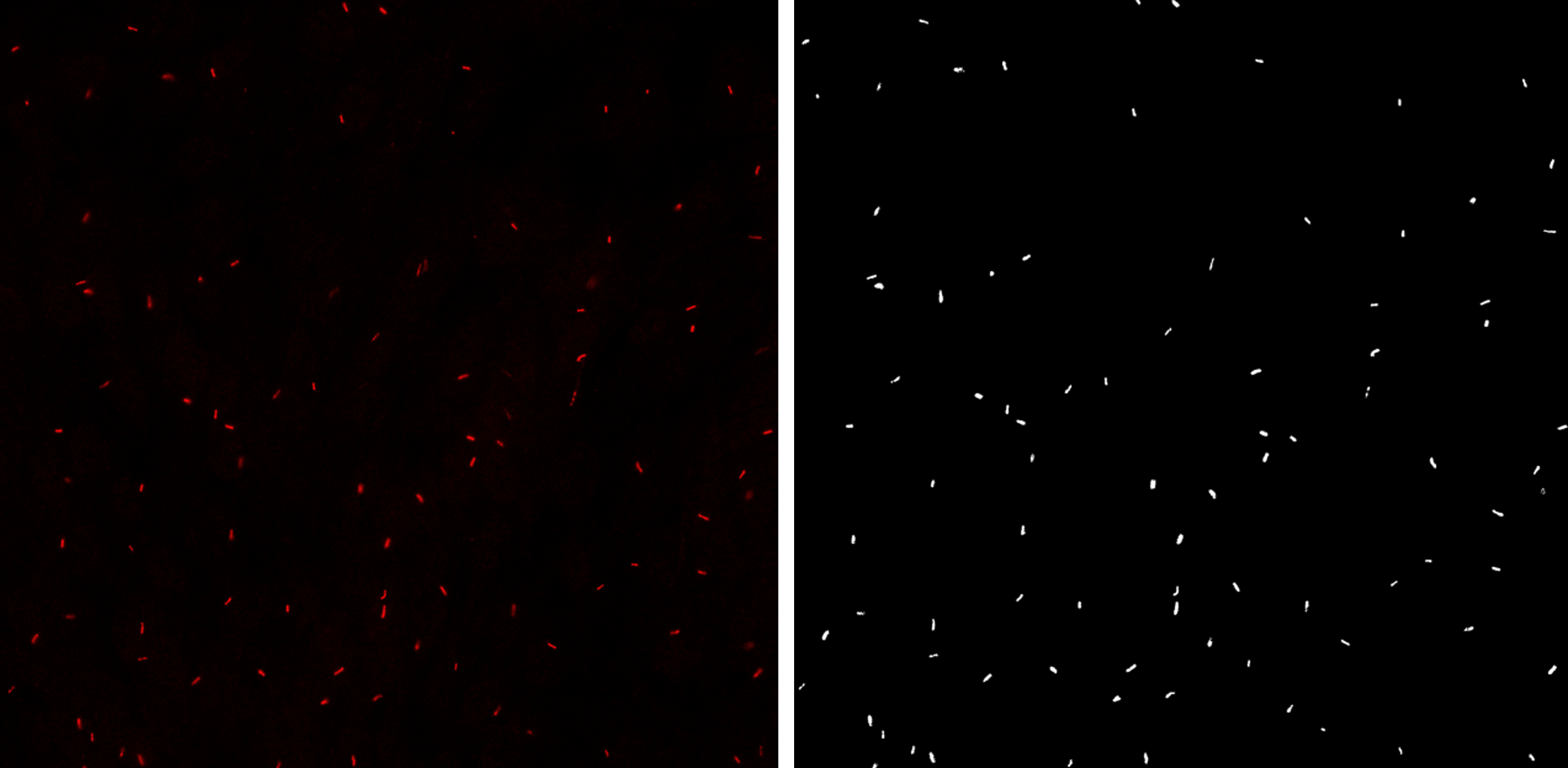
Cilia
Cilia mask